


DNA Structure and Shape
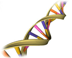
DNA, also known, as deoxyribonucleic acid is a molecule, which is a bunch of atoms stuck together. In the case of DNA, these atoms are combined to form the shape of a long spiraling ladder. We can see the picture here clearly to recognize the shape of the DNA.
If you ever studied biology, you probably heard that DNA acts as a blueprint or recipe for living thing. How on earth can a mere molecule act as a blueprint for something as complex and wonderful as a tree, a dog and human beings? That’s really amazing.
DNA is one of the ultimate instruction guides. It is more complex than any how-to book you have ever used. The entire instruction guide is written in code. If you look at the chemical structure of DNA closely, it will show four main building blocks. We call these nitrogenous bases: Adenine (A), Thymine (T), Guanine (G), and Cytosine (C). DNA also includes sugars and phosphate groups (made of phosphorus and oxygen). These make the phosphate-deoxyribose backbone.
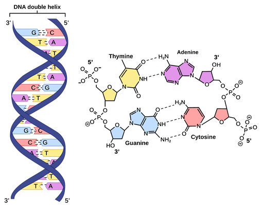
If you think of the structure of DNA as a ladder, the rungs of the ladder are made from the nitrogenous bases. These bases pair up to make each step of the ladder. They also only pair up in a specific way. (A) always pairs with (T) and (G) always pairs with (C). This is very important when it is time to copy all or part of the DNA.
So, to answer the question, what is DNA? DNA is a molecular blueprint for a living thing. DNA creates RNA, and RNA creates protein, and proteins go on to form life. This entire process is complicated, sophisticated and magical and it is entirely based in chemistry that can be studied and understood.
How to Separate DNA Fragment?
AS we said that DNA can be studied and understood, but how can we do it? The scientists learn and research and explore them. People use gel electrophoresis to separate DNA for further research. Gel electrophoresis is a technique used to separate DNA fragments (or other macromolecules, such as RNA and proteins) based on their size and charge. Electrophoresis involves running a current through a gel containing the molecules of interest. Based on their size and charge, the molecules will travel through the gel in different directions or at different speeds, allowing them to be separated from one another. Using electrophoresis, we can see how many different DNA fragments are present in a sample and how large they are relative to one another.
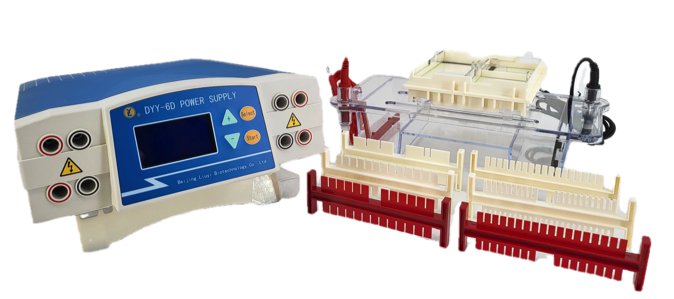
If you want to do the gel electrophoresis, first you need the related experimental equipment, the electrophoresis cell (tank/chamber) and its power supply. The following picture shows a horizontal electrophoresis cell (tank/chamber) the model DYCP-31DN and the power supply the model DYY-6D from Beijing Liuyi Biotechnology Co., Ltd for DNA gel electrophoresis.
Gel electrophoresis involves gel, which is kind of Jello-like material. Gels for DNA separation are often used agarose, which comes as dry, powdered flakes. When the agarose is heated in a buffer (water with some salts in it) and allowed to cool, it will form a solid, slightly squishy gel. At the molecular level, the gel is a matrix of agarose molecules that are held together by hydrogen bonds and form tiny pores.
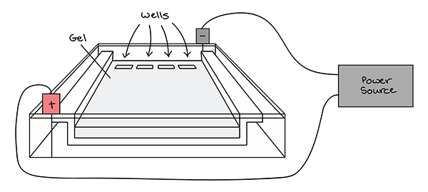
Image from Khan Academy
After preparation the gel, put the gel in the electrophoresis cell’s tank body, and pour buffer solution in the buffer tank until get immersed the gel. Then DNA samples are loaded into wells (indentations) at one end of a gel, and an electric current is applied to pull them through the gel. DNA fragments are negatively charged, so they move towards the positive electrode. Because all DNA fragments have the same amount of charge per mass, small fragments move through the gel faster than large ones. After running gel electrophoresis, the DNA fragments have been separated; and the researchers can examine the gel and see what sizes of bands are found on it. When a gel is stained with a DNA-binding dye and placed under UV light, the DNA fragments will glow, allowing us to see the DNA present at different locations along the length of the gel.
Except electrophoresis cells (tanks/chambers) and power supplies, Beijing Liuyi Biotechnology Co., Ltd also provides UV transilluminator, which can observe and take photos for protein and DNA electrophoresis gel. The model WD-9403B is a portable UV transilluminator for observing DNA electrophoresis gel. The model WD-9403F can observe, take photos for both protein and DNA gel.
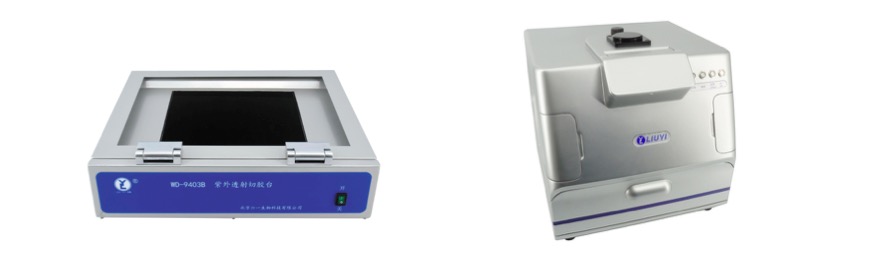
WD-9403B
WD-9403F
Beijing Liuyi Biotechnology Co., Ltd has more than 50 years history in China and it can provide stable and high-quality products all around the world. Through years’ development, it is worthy of your choice!
For more information about us, please contact us by email sales@ly.com.cn or sales01@ly.com.cn.
Post time: May-13-2022


